
Pectus Excavatum, often abbreviated as PE, is a congenital chest wall deformity characterized by a sunken appearance of the sternum or breastbone. This condition can range from mild to severe and may affect individuals both physically and emotionally. While it is not life-threatening in most cases, understanding its causes, symptoms, diagnostic methods, and treatment options is crucial for those affected by it. In this article, we will delve into the details of Pectus Excavatum, providing comprehensive insights into the condition.
What is Pectus Excavatum?
Pectus Excavatum is a structural abnormality where the breastbone appears to be caved inward, giving the chest a concave or “funnel-like” shape. It is one of the most common chest wall deformities, occurring in approximately 1 in every 300 to 400 births. The severity of the condition varies widely, with some individuals experiencing only a slight indentation, while others may have a more pronounced deformity that impacts heart and lung function.
Physical Appearance
- A noticeable dip in the center of the chest
- The breastbone may press against the heart or lungs in severe cases
- The shoulders may appear rounded or hunched forward
Impact on Health
- Mild cases may cause no physical symptoms but could lead to self-consciousness
- Severe cases may compress the heart or lungs, leading to reduced exercise tolerance or shortness of breath
- In rare instances, it may contribute to heart murmurs or arrhythmias
Causes of Pectus Excavatum
The exact cause of Pectus Excavatum is not fully understood, but researchers believe it results from a combination of genetic and developmental factors. Below are some of the potential causes and contributing factors:
Genetic Factors
Many individuals with Pectus Excavatum have a family history of the condition, suggesting a genetic component. Studies indicate that certain gene mutations may predispose individuals to chest wall abnormalities. If a close family member has the condition, the likelihood of developing it increases.
Abnormal Growth of Cartilage
The chest wall is made up of bones and cartilage. During fetal development and early childhood, abnormal growth or overgrowth of the cartilage connecting the ribs to the breastbone can lead to the inward curvature characteristic of this condition.
Connective Tissue Disorders
Some connective tissue disorders, such as Marfan syndrome or Ehlers-Danlos syndrome, are associated with an increased risk of chest wall deformities. These conditions affect the body’s connective tissues, which play a critical role in maintaining the structure of the chest wall.
Environmental Influences
While less commonly discussed, environmental factors during pregnancy or early childhood may also contribute to the development of Pectus Excavatum. For instance, nutritional deficiencies or exposure to certain toxins could potentially influence fetal development.
Symptoms and Complications
Pectus Excavatum does not always present with obvious symptoms, especially in mild cases. However, as the condition progresses or becomes more severe, certain physical and psychological symptoms may arise.
Physical Symptoms
- Shortness of breath, particularly during physical activity
- Fatigue or reduced stamina
- Rapid heart rate or palpitations
- Chest pain or discomfort, though this is less common
Psychological and Emotional Impact
- Low self-esteem due to the visible deformity
- Social anxiety or withdrawal
- Body image issues, particularly during adolescence
Potential Complications
In severe cases, the inward curvature of the breastbone can press against the heart and lungs, leading to complications such as:
- Reduced lung capacity
- Heart murmurs or mitral valve prolapse
- Exercise intolerance
Diagnosing Pectus Excavatum
Diagnosing this condition typically involves a combination of physical examination, imaging tests, and sometimes functional assessments. Early diagnosis is important to determine the severity of the condition and plan appropriate treatment.
Physical Examination
A healthcare provider will begin by examining the chest visually and manually. They may ask the patient to lie flat on their back or perform simple movements to assess the extent of the deformity.
Imaging Tests
- Chest X-ray: Provides a basic view of the chest structure and helps identify abnormalities.
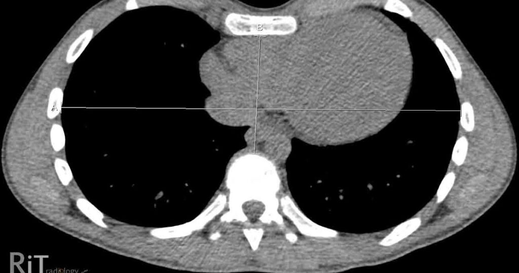
- CT Scan: Offers a detailed cross-sectional image of the chest, allowing doctors to measure the severity of the deformity using a standardized index called the Haller Index.
- MRI: Useful for evaluating the impact of the deformity on the heart and lungs without exposing the patient to radiation.
Pulmonary Function Tests
These tests measure how well the lungs are functioning. They can help determine if the deformity is affecting breathing capacity.
Cardiac Evaluation
In cases where the heart is suspected to be compressed, an echocardiogram may be performed to assess heart function and detect any abnormalities such as valve issues or reduced cardiac output.
Treatment Options for Pectus Excavatum
The treatment approach for Pectus Excavatum depends on the severity of the condition, the presence of symptoms, and the individual’s age. While mild cases may not require intervention, moderate to severe cases often benefit from medical or surgical treatments.
Non-Surgical Treatments
For individuals with mild deformities or those who are not candidates for surgery, non-surgical options may be recommended:
- Vacuum Bell Therapy: A non-invasive device that uses suction to gradually lift the breastbone outward over time. It is most effective in children and adolescents whose bones are still growing.
- Physical Therapy: Exercises designed to strengthen the chest muscles and improve posture may help alleviate some symptoms and improve appearance.
- Bracing: Custom braces can be used in younger patients to correct the deformity as they grow.
Surgical Treatments
Surgery is typically reserved for severe cases or when the deformity significantly impacts heart and lung function. Two main surgical procedures are commonly performed:
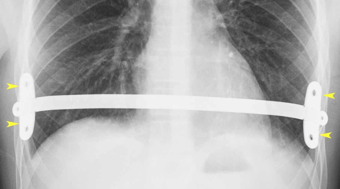
- Nuss Procedure: A minimally invasive technique where a curved metal bar is inserted under the sternum to push it outward. The bar remains in place for two to three years before being removed.
- Ravitch Procedure: An open surgery that involves removing abnormal cartilage and reshaping the chest wall. This procedure is more invasive and is usually considered for older patients or those with complex deformities.
Post-Surgical Care
After surgery, patients require careful monitoring and follow-up care to ensure proper healing. Pain management, physical therapy, and restrictions on physical activities are typically part of the recovery process.
Lifestyle Adjustments and Support
Beyond medical treatments, individuals with Pectus Excavatum may benefit from making lifestyle adjustments and seeking emotional support:
- Exercise: Low-impact activities like swimming or yoga can help improve overall fitness without straining the chest.
- Counseling: Speaking with a therapist or joining a support group can help address emotional challenges related to body image and self-esteem.
- Educational Resources: Learning about the condition through reputable sources can empower patients and their families to make informed decisions about care.
When to Seek Medical Advice
If you or your child exhibits signs of Pectus Excavatum, it is important to consult a healthcare professional. Early evaluation can help determine whether the condition requires intervention and rule out other underlying health issues. Seek immediate medical attention if symptoms such as severe chest pain, difficulty breathing, or rapid heartbeat occur.
Research and Advances in Treatment
Ongoing research continues to explore new techniques and technologies for managing Pectus Excavatum. Innovations in minimally invasive surgeries, improved vacuum bell designs, and advancements in genetic studies hold promise for better outcomes in the future. Staying informed about these developments can help patients and families make proactive decisions about their care.


