
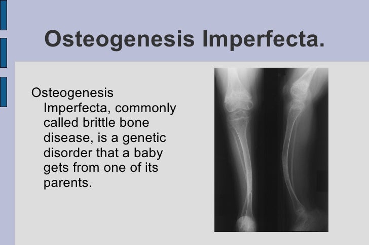
Osteogenesis Imperfecta, commonly abbreviated as OI, is a rare genetic disorder characterized by fragile bones that break easily. Often referred to as brittle bone disease, this condition affects the body’s ability to produce strong and healthy bones. While the severity of the disease varies widely among individuals, it poses significant challenges for those living with it. In this article, we will explore the causes, symptoms, types, diagnosis, treatment options, and ongoing research related to this complex condition.
Understanding the Causes of Brittle Bone Disease
The primary cause of brittle bone disease lies in genetic mutations that affect collagen production. Collagen is a protein that serves as a crucial building block for bones, skin, and connective tissues. When there are defects in the genes responsible for collagen production, the bones become weak and prone to fractures. These genetic mutations can be inherited from one or both parents or may occur spontaneously during fetal development.
In most cases, the mutations occur in the COL1A1 or COL1A2 genes, which provide instructions for making type I collagen. Type I collagen is the most abundant form of collagen in the human body and plays a vital role in maintaining bone strength. When these genes are defective, the collagen produced is either insufficient or of poor quality, leading to the characteristic fragility of bones seen in this condition.
Types of Brittle Bone Disease
Brittle bone disease is classified into several types based on the severity of symptoms and the specific genetic mutations involved. Each type presents unique challenges and requires tailored approaches to management and care.
Type I: Mild Form
- This is the mildest and most common form of brittle bone disease.
- Individuals with this type typically experience fewer fractures compared to other forms.
- Bone deformities are usually minimal, and height is often normal or slightly below average.
- Hearing loss may develop later in life due to abnormalities in the ear bones.
Type II: Severe and Often Fatal
- This is the most severe form and is often fatal shortly after birth.
- Babies born with this type have extremely fragile bones, leading to multiple fractures even before birth.
- Underdeveloped lungs and respiratory complications contribute to the high mortality rate.
Type III: Progressive Deformities
- This type is characterized by progressive bone deformities and frequent fractures.
- Children with this form often have short stature and may require mobility aids such as wheelchairs.
- Scoliosis (curvature of the spine) and other skeletal abnormalities are common.
Type IV: Moderate Severity
- This type falls between mild and severe forms in terms of severity.
- Fractures occur more frequently than in Type I but less often than in Type III.
- Individuals may experience some degree of short stature and bone deformities.
Recognizing the Symptoms
The symptoms of brittle bone disease vary depending on the type and severity of the condition. However, some common signs and symptoms include:
- Frequent bone fractures, often with minimal trauma or no apparent cause.
- Bone deformities, such as bowed legs or curved spine.
- Short stature due to impaired bone growth.
- Blue sclerae, where the whites of the eyes appear bluish due to thinness of the eye tissue.
- Hearing loss, particularly in adults with Type I.
- Dental issues, such as brittle teeth prone to cavities and breakage.
- Muscle weakness and joint laxity.
Diagnosis of Brittle Bone Disease
Diagnosing brittle bone disease involves a combination of clinical evaluation, imaging studies, and genetic testing. Early diagnosis is crucial for implementing appropriate management strategies and improving quality of life.
Clinical Evaluation
- Doctors assess the patient’s medical history, including family history of fractures or similar conditions.
- A physical examination helps identify signs such as blue sclerae, short stature, and bone deformities.
Imaging Studies
- X-rays are commonly used to detect fractures, bone deformities, and changes in bone density.
- Dual-energy X-ray absorptiometry (DEXA) scans may be performed to measure bone mineral density.
Genetic Testing
- Genetic tests analyze the DNA to identify mutations in the COL1A1, COL1A2, or other related genes.
- This helps confirm the diagnosis and determine the specific type of brittle bone disease.
Treatment Options and Management Strategies
While there is no cure for brittle bone disease, various treatment options aim to manage symptoms, prevent fractures, and improve overall quality of life. A multidisciplinary approach involving healthcare professionals from different specialties is often necessary.
Medical Interventions
Several medications and therapies are available to strengthen bones and reduce fracture risk:
- Bisphosphonates: These drugs help increase bone density by slowing down bone resorption. They are commonly prescribed for children and adults with brittle bone disease.
- Growth Hormone Therapy: This treatment may be used to promote bone growth in children with certain types of the condition.
- Pain Management: Pain relievers and anti-inflammatory medications are prescribed to manage pain associated with fractures and chronic discomfort.
Physical and Occupational Therapy
Therapy plays a critical role in improving mobility, strength, and independence:
- Physical therapy focuses on strengthening muscles, improving balance, and enhancing mobility.
- Occupational therapy helps individuals adapt to daily activities and use assistive devices effectively.
Surgical Interventions
In some cases, surgery may be necessary to address complications associated with brittle bone disease:
- Rodding surgery involves inserting metal rods into long bones to stabilize them and prevent fractures.
- Spinal surgeries may be performed to correct scoliosis or other spinal deformities.
Lifestyle Modifications
Making certain lifestyle adjustments can significantly reduce the risk of fractures and improve overall well-being:
- Avoiding high-impact activities and sports that pose a risk of injury.
- Maintaining a balanced diet rich in calcium and vitamin D to support bone health.
- Using protective gear, such as braces or helmets, to minimize injury risks.
Ongoing Research and Future Directions
Research into brittle bone disease continues to advance, offering hope for improved treatments and potential cures. Scientists are exploring innovative approaches such as gene therapy, stem cell therapy, and novel drug compounds to target the underlying genetic causes of the condition.
Gene Therapy
Gene therapy aims to correct defective genes responsible for collagen production. By introducing functional copies of the affected genes, researchers hope to restore normal collagen synthesis and improve bone strength.
Stem Cell Therapy
Stem cells have the potential to develop into various cell types, including bone-forming cells. Researchers are investigating how stem cells can be used to regenerate damaged bone tissue and enhance bone healing.
New Drug Development
Pharmaceutical companies are working on developing drugs that target specific pathways involved in bone formation and remodeling. These drugs could offer more effective and targeted treatments for individuals with brittle bone disease.
Collaborative Efforts
International collaborations and patient registries are playing a vital role in advancing research. By collecting data from individuals with brittle bone disease, scientists can better understand the condition’s natural history and identify patterns that inform future studies.


