Lung nodules, also known as pulmonary nodules, are small, abnormal growths or masses that form in the lungs. These growths are often detected incidentally during imaging tests like chest X-rays or CT scans performed for unrelated reasons. While many lung nodules are benign, some may indicate an underlying condition such as cancer or infection. Understanding lung nodules is crucial for early detection and appropriate management. In this article, we will explore what lung nodules are, their types, how they are diagnosed, and the available treatment options.
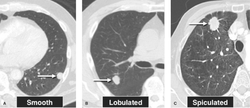
What Are Lung Nodules?
Lung nodules are defined as round or oval-shaped growths within the lung tissue that measure less than three centimeters in diameter. They can appear as solitary lesions or multiple growths scattered throughout the lungs. Most lung nodules do not cause noticeable symptoms, which is why they are often discovered accidentally during routine medical examinations or diagnostic procedures for other health concerns.
The causes of lung nodules vary widely. Some are benign and result from inflammation, scarring, or infections, while others may be malignant and represent early-stage lung cancer or metastatic disease. Given their potential significance, it is essential to evaluate lung nodules thoroughly to determine their nature and whether further intervention is necessary.
Common Causes of Lung Nodules
- Infections such as tuberculosis or fungal diseases
- Inflammatory conditions like sarcoidosis or rheumatoid arthritis
- Previous lung injuries or scars from healed infections
- Noncancerous growths such as hamartomas
- Malignant tumors originating in the lungs or spreading from other parts of the body
Types of Lung Nodules
Lung nodules are broadly categorized into two main types based on their composition and appearance on imaging studies: solid nodules and subsolid nodules. Each type has distinct characteristics that help doctors assess their likelihood of being benign or malignant.
Solid Nodules
Solid nodules are dense, well-defined growths that appear uniformly opaque on imaging tests. They are the most common type of lung nodule and can result from a variety of causes, including benign conditions like scars and more serious issues like cancer. The risk of malignancy in solid nodules depends on factors such as size, shape, and growth rate over time.
Subsolid Nodules
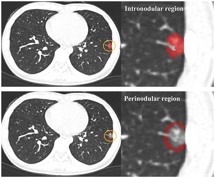
Subsolid nodules are less dense than solid nodules and have a hazy or ground-glass appearance on imaging studies. This category includes two subtypes: pure ground-glass nodules and part-solid nodules. Pure ground-glass nodules consist entirely of hazy tissue, while part-solid nodules contain both ground-glass and solid components. Subsolid nodules are more likely to be associated with malignancy compared to solid nodules, making them a focus of careful monitoring and evaluation.
Diagnosis of Lung Nodules
Diagnosing lung nodules involves a combination of imaging studies, clinical evaluation, and sometimes invasive procedures. The goal is to determine whether a nodule is benign or requires further investigation for potential malignancy. Here is an overview of the diagnostic process:
Imaging Studies
The initial step in diagnosing lung nodules is identifying them through imaging tests. Chest X-rays and computed tomography scans are the primary tools used for this purpose. Computed tomography provides detailed images of the lungs and allows doctors to assess the size, shape, location, and density of nodules with greater precision than X-rays.
Follow-up imaging may be recommended to monitor changes in the nodule over time. For example, if a nodule remains stable in size and appearance over several months or years, it is more likely to be benign. Conversely, rapid growth or changes in the nodule’s structure may raise suspicion for malignancy.
Risk Assessment
Doctors use various risk assessment tools to evaluate the likelihood of a lung nodule being cancerous. Factors considered include the patient’s age, smoking history, family history of lung cancer, presence of symptoms, and specific features of the nodule observed on imaging studies. One commonly used tool is the Brock University model, which assigns a probability score based on these variables.
Biopsy and Further Testing
If a lung nodule is deemed suspicious for malignancy, additional testing may be required to confirm the diagnosis. A biopsy involves removing a small sample of tissue from the nodule for microscopic examination. This can be done using minimally invasive techniques such as bronchoscopy, where a thin tube is inserted through the airways, or percutaneous needle biopsy, where a needle is guided through the skin into the lung.
In some cases, advanced imaging techniques like positron emission tomography scans may be used to assess metabolic activity within the nodule. High metabolic activity is often associated with cancerous growths.
Treatment Options for Lung Nodules
The treatment approach for lung nodules depends on whether they are benign or malignant, as well as the patient’s overall health and preferences. Below are the main strategies used to manage lung nodules:
Observation and Monitoring
For nodules that are highly likely to be benign, a “watchful waiting” approach is often recommended. This involves regular follow-up imaging at specified intervals to monitor for any changes in the nodule’s size or appearance. If the nodule remains stable over time, no further intervention may be needed.
Surgical Removal
If a nodule is suspected or confirmed to be malignant, surgical removal may be necessary. Procedures such as wedge resection, lobectomy, or pneumonectomy may be performed depending on the size and location of the nodule. Minimally invasive techniques like video-assisted thoracoscopic surgery are often preferred due to their shorter recovery times and reduced complications.
Radiation Therapy
Radiation therapy may be used as an alternative to surgery for patients who are not good candidates for surgical intervention. Stereotactic body radiation therapy delivers high doses of targeted radiation to destroy cancerous cells while sparing surrounding healthy tissue.
Targeted Therapy and Immunotherapy
For malignant nodules that are part of advanced lung cancer, systemic treatments such as targeted therapy and immunotherapy may be employed. Targeted therapies block specific molecules involved in cancer growth, while immunotherapy enhances the body’s immune response against cancer cells.
Treatment for Benign Nodules
Benign nodules typically do not require aggressive treatment unless they cause symptoms or complications. In rare cases where a benign nodule leads to breathing difficulties or recurrent infections, surgical removal or other interventions may be considered.
Prevention and Lifestyle Considerations
While not all lung nodules can be prevented, certain lifestyle modifications can reduce the risk of developing malignant nodules. Quitting smoking is one of the most effective ways to lower the risk of lung cancer and other respiratory conditions. Avoiding exposure to environmental pollutants, asbestos, and radon gas is also important for maintaining lung health.
Regular check-ups and screenings are recommended for individuals at higher risk of developing lung nodules, such as those with a history of smoking or significant occupational exposures. Early detection through screening programs like low-dose computed tomography scans can improve outcomes by identifying nodules before they progress to advanced stages.
Emotional and Psychological Support
Being diagnosed with a lung nodule can be a stressful experience, especially when there is uncertainty about its nature. Patients may benefit from emotional and psychological support during the diagnostic and treatment process. Counseling, support groups, and educational resources can help individuals cope with anxiety and make informed decisions about their care.
