
A hydatidiform mole, often abbreviated as HM, is a rare and abnormal growth that occurs in the uterus during early pregnancy. This condition results in the formation of cysts that resemble clusters of grapes, hence its descriptive nickname. While it may sound alarming, understanding this condition can help individuals recognize symptoms early and seek appropriate medical care. In this article, we will explore the nature of this condition, its causes, how it is diagnosed, and the available treatment options.
What is a Hydatidiform Mole?
A hydatidiform mole is a type of gestational trophoblastic disease, which refers to a group of conditions that involve abnormal growth of cells in the tissue that would normally develop into the placenta. Instead of forming a healthy fetus, the cells grow uncontrollably, leading to the development of fluid-filled sacs or cysts. These cysts give the uterine tissue a grape-like appearance when examined under a microscope.
There are two main types of hydatidiform moles:
- Complete Hydatidiform Mole: In this type, no normal fetal tissue develops. The entire growth consists of abnormal cells that form cysts.
- Partial Hydatidiform Mole: In this case, some fetal tissue may be present alongside the abnormal growth. However, the fetus is usually not viable.
Causes of a Hydatidiform Mole
The exact cause of a hydatidiform mole is related to chromosomal abnormalities that occur during fertilization. Normally, a fertilized egg contains 46 chromosomes—23 from the mother and 23 from the father. In cases of a hydatidiform mole, this balance is disrupted due to errors in cell division.
Complete Hydatidiform Mole
In a complete hydatidiform mole, all of the chromosomes come from the father. This happens when an empty egg, lacking maternal chromosomes, is fertilized by one or two sperm cells. The resulting cells have only paternal chromosomes, which leads to uncontrolled growth and the absence of normal fetal development.
Partial Hydatidiform Mole
In a partial hydatidiform mole, there is an imbalance in the number of chromosomes contributed by each parent. Typically, the egg is fertilized by two sperm cells, resulting in an extra set of paternal chromosomes. This genetic abnormality causes both abnormal tissue growth and the presence of some fetal tissue, though the fetus is usually not viable.
Risk factors for developing a hydatidiform mole include:
- Maternal age: Women who are younger than 20 or older than 35 are at a higher risk.
- Previous history: Women who have had a hydatidiform mole in the past are more likely to experience it again.
- Dietary deficiencies: A diet low in certain nutrients, such as vitamin A or folic acid, may increase the risk.
Symptoms of a Hydatidiform Mole
Recognizing the symptoms of a hydatidiform mole is crucial for early diagnosis and treatment. Some common signs and symptoms include:
- Vaginal bleeding during the first trimester, which may contain small, grape-like cysts.
- Severe nausea and vomiting, often more intense than typical morning sickness.
- Rapid uterine growth, where the uterus becomes larger than expected for the stage of pregnancy.
- Pelvic pain or pressure caused by the abnormal growth.
- High levels of human chorionic gonadotropin, a hormone produced during pregnancy, which may indicate abnormal cell growth.
In some cases, women with a hydatidiform mole may not experience any noticeable symptoms until routine prenatal testing reveals abnormalities.
Diagnosis of a Hydatidiform Mole
Diagnosing a hydatidiform mole involves a combination of physical examinations, laboratory tests, and imaging studies. Early detection is essential to prevent complications and ensure timely treatment.
Physical Examination
A healthcare provider may notice unusual findings during a pelvic exam, such as an abnormally enlarged uterus or vaginal bleeding. These observations can prompt further investigation.
Laboratory Tests
Blood tests are used to measure levels of human chorionic gonadotropin. Elevated levels of this hormone, especially when they do not correspond to the expected stage of pregnancy, may suggest the presence of a hydatidiform mole.
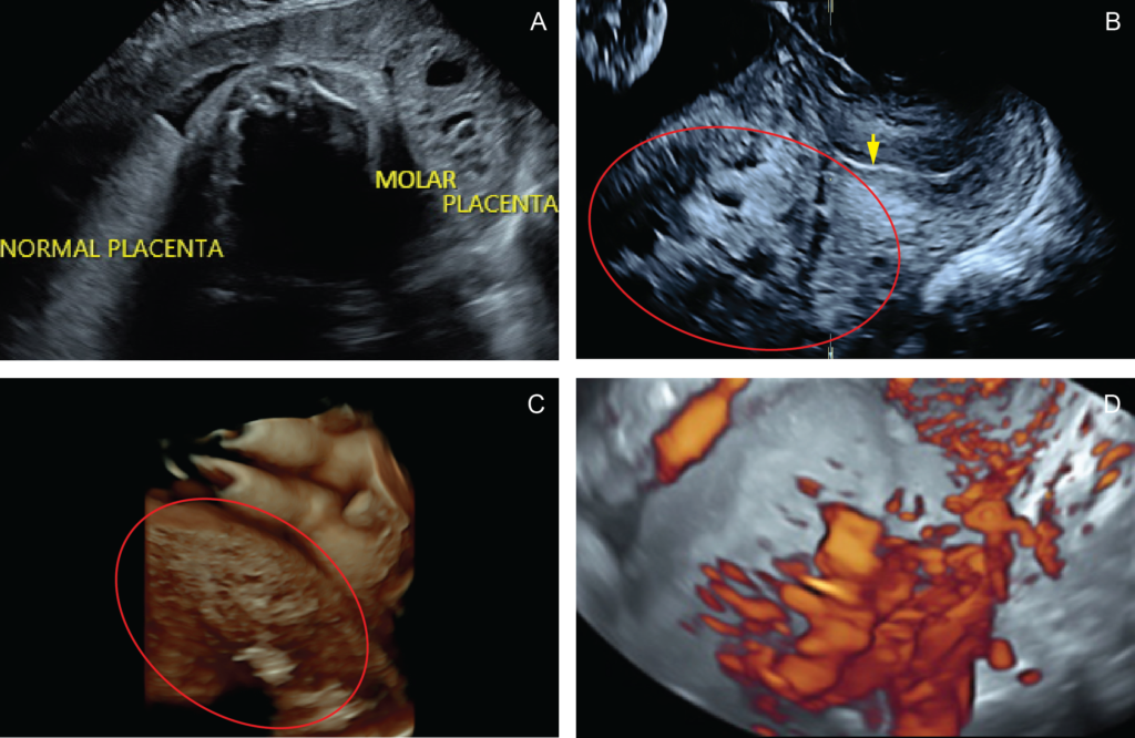
Ultrasound Imaging
An ultrasound is a key diagnostic tool for identifying a hydatidiform mole. During the procedure, the ultrasound technician looks for characteristic features, such as the absence of a viable fetus, the presence of cystic spaces within the uterine tissue, and an abnormally thickened placenta. These findings help confirm the diagnosis.
Histopathological Examination
If a hydatidiform mole is suspected, the tissue removed during a surgical procedure is sent to a laboratory for examination under a microscope. This histopathological analysis confirms the diagnosis by identifying the presence of abnormal cells and cysts.
Treatment Options for a Hydatidiform Mole
Treatment for a hydatidiform mole focuses on removing the abnormal tissue and monitoring for potential complications. The approach depends on the type of mole, the patient’s overall health, and whether there are any signs of persistent disease.
Surgical Removal
The primary treatment for a hydatidiform mole is a procedure called dilation and curettage. During this surgery, the cervix is dilated, and the abnormal tissue is gently scraped from the uterine lining. This procedure is typically performed under general anesthesia and is highly effective in removing the growth.
Monitoring After Treatment
After the removal of a hydatidiform mole, patients are closely monitored to ensure that all abnormal cells have been eliminated. Regular blood tests are conducted to track levels of human chorionic gonadotropin. If these levels remain elevated or begin to rise again, it may indicate that some abnormal tissue remains or has progressed to a more serious condition, such as gestational trophoblastic neoplasia.
Chemotherapy
In cases where abnormal cells persist or spread beyond the uterus, chemotherapy may be required. Chemotherapy uses medications to target and destroy rapidly dividing cells, including those associated with a hydatidiform mole. The specific drugs and duration of treatment depend on the severity of the condition and the patient’s response to therapy.
Hysterectomy
For women who do not wish to have children in the future, a hysterectomy may be considered. This surgical procedure involves the removal of the uterus and is typically reserved for cases where other treatments have failed or when the patient prefers a permanent solution.
Potential Complications
If left untreated, a hydatidiform mole can lead to serious complications. One of the most significant risks is the development of gestational trophoblastic neoplasia, a malignant condition that requires more aggressive treatment. Other potential complications include:
- Excessive bleeding during or after the removal of the mole.
- Infection of the uterine tissue.
- Preeclampsia, a condition characterized by high blood pressure and organ damage, which can occur even in the absence of a viable pregnancy.
Emotional and Psychological Impact
Experiencing a hydatidiform mole can be emotionally challenging for many women and their families. The loss of a pregnancy, combined with concerns about future fertility and health, can lead to feelings of grief, anxiety, and uncertainty. It is important for patients to seek support from healthcare providers, counselors, or support groups to address these emotional needs.
Prevention and Future Pregnancies
While it is not always possible to prevent a hydatidiform mole, certain measures can reduce the risk. Maintaining a balanced diet rich in essential nutrients, such as folic acid and vitamin A, may help lower the likelihood of developing this condition. Additionally, women with a history of hydatidiform moles should consult their healthcare provider before attempting to conceive again.
Most women who have had a hydatidiform mole can go on to have healthy pregnancies in the future. However, close monitoring during subsequent pregnancies is recommended to ensure early detection of any abnormalities.


