
Enlarged veins in the esophagus, commonly referred to as esophageal varices, are a serious medical condition that can lead to life-threatening complications if left untreated. These swollen veins develop in the lining of the esophagus, often as a result of increased pressure in the portal vein system. The portal vein carries blood from the digestive organs to the liver, and when this system is disrupted, it can cause blood to back up into smaller vessels, including those in the esophagus. This article provides an in-depth look at esophageal varices, exploring their causes, symptoms, diagnostic methods, and available treatments.
Understanding Esophageal Varices
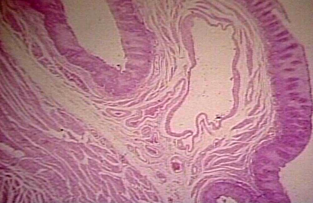
Esophageal varices occur when the veins in the esophagus become abnormally enlarged due to increased pressure within the portal venous system. The portal vein is responsible for transporting nutrient-rich blood from the intestines, spleen, and pancreas to the liver. When the flow of blood through the liver is obstructed or slowed down, the pressure within the portal vein rises, leading to a condition known as portal hypertension. As a result, blood seeks alternative pathways to bypass the liver, causing smaller veins in the esophagus to swell and become varices.
These swollen veins are fragile and prone to rupture, which can result in severe bleeding. Bleeding from esophageal varices is a medical emergency and requires immediate attention. Understanding the underlying mechanisms and risk factors associated with this condition is crucial for early detection and effective management.
Causes of Enlarged Veins in the Esophagus
The primary cause of esophageal varices is portal hypertension, which itself can arise from various underlying conditions. Below are some of the most common causes:
- Liver Cirrhosis: Liver cirrhosis is the leading cause of portal hypertension. It occurs when healthy liver tissue is replaced by scar tissue, often due to chronic alcohol consumption, viral hepatitis, or non-alcoholic fatty liver disease. The scarring disrupts the normal blood flow through the liver, increasing pressure in the portal vein.
- Blood Clots: Blood clots in the portal vein or its branches can obstruct blood flow, leading to increased pressure and the development of varices.
- Schistosomiasis: This parasitic infection, common in certain parts of the world, can damage the liver and lead to portal hypertension.
- Budd-Chiari Syndrome: This rare condition involves the blockage of the hepatic veins, which drain blood from the liver. The obstruction can cause portal hypertension and subsequent varices.
- Other Liver Diseases: Conditions such as primary biliary cirrhosis and autoimmune hepatitis can also contribute to portal hypertension and the formation of esophageal varices.
Risk Factors for Developing Esophageal Varices
Certain individuals are at a higher risk of developing esophageal varices due to predisposing factors. These include:
- A history of chronic liver disease or cirrhosis
- Prolonged alcohol abuse
- Infection with hepatitis B or hepatitis C viruses
- Obesity and metabolic syndrome
- Genetic predisposition to liver diseases
Symptoms of Esophageal Varices
Esophageal varices often do not cause noticeable symptoms until they rupture and bleed. However, some individuals may experience warning signs before a major bleeding episode. These symptoms include:
- Vomiting blood, which may appear bright red or resemble coffee grounds
- Black, tarry stools caused by the presence of digested blood
- Dizziness or lightheadedness due to blood loss
- Rapid heart rate and low blood pressure
- Confusion or altered mental state, particularly in cases of significant blood loss
If any of these symptoms occur, it is essential to seek medical attention immediately, as they may indicate a life-threatening situation.
Diagnosing Esophageal Varices
Diagnosing esophageal varices typically involves a combination of clinical evaluation, imaging tests, and endoscopic procedures. Here are the most common diagnostic methods used:
- Endoscopy: An upper gastrointestinal endoscopy is the gold standard for diagnosing esophageal varices. During this procedure, a flexible tube with a camera is inserted through the mouth to examine the esophagus and stomach. The presence, size, and severity of varices can be assessed during the examination.
- Imaging Tests: Imaging techniques such as ultrasound, CT scans, or MRI may be used to evaluate the liver and portal vein system. These tests can help identify underlying causes of portal hypertension and assess the extent of liver damage.
- Laboratory Tests: Blood tests may be performed to check liver function, detect infections, and measure clotting times. Abnormal results can provide clues about the presence of liver disease or other contributing factors.
Treatment Options for Esophageal Varices
The treatment of esophageal varices focuses on two main objectives: preventing bleeding and managing underlying conditions that contribute to portal hypertension. Depending on the severity of the varices and the patient’s overall health, different treatment approaches may be employed.
Preventing Bleeding
For individuals with large or high-risk varices, preventive measures are essential to reduce the likelihood of bleeding. These include:
- Beta-Blockers: Medications such as propranolol and nadolol are commonly prescribed to lower blood pressure in the portal vein system. By reducing the pressure, these drugs decrease the risk of variceal rupture.
- Endoscopic Band Ligation: This minimally invasive procedure involves placing small rubber bands around the varices during an endoscopy. The bands cut off blood flow to the varices, causing them to shrink and eventually disappear.
Managing Acute Bleeding
If esophageal varices bleed, prompt intervention is necessary to control the hemorrhage and stabilize the patient. Treatment options for acute bleeding include:
- Endoscopic Therapy: Endoscopic band ligation or sclerotherapy may be performed to stop active bleeding. Sclerotherapy involves injecting a solution directly into the varices to cause them to collapse and seal off.
- Medications: Vasopressin and octreotide are medications that can be administered to constrict blood vessels and reduce bleeding.
- Transjugular Intrahepatic Portosystemic Shunt (TIPS): In severe cases, a TIPS procedure may be performed. This involves creating a shunt between the portal vein and hepatic vein to relieve pressure in the portal system.
Addressing Underlying Liver Disease
Since liver disease is the primary cause of portal hypertension and esophageal varices, managing the underlying condition is critical for long-term success. Treatment strategies may include:
- Lifestyle modifications such as abstaining from alcohol and maintaining a healthy diet
- Antiviral medications for hepatitis B or hepatitis C infections
- Management of complications such as ascites, hepatic encephalopathy, and malnutrition
- In advanced cases, liver transplantation may be considered as a definitive treatment option
Monitoring and Follow-Up
Regular monitoring is essential for individuals diagnosed with esophageal varices, even if they have not experienced bleeding. Follow-up endoscopies are typically recommended every one to two years to assess the status of the varices and determine the need for additional interventions. Patients should also work closely with their healthcare providers to manage risk factors and optimize their overall health.


