
A cholesteatoma is a serious medical condition that requires prompt attention. Often abbreviated as CKT in medical literature, this abnormal growth of skin cells in the middle ear can lead to significant complications if left untreated. This article delves into the causes, symptoms, diagnostic methods, and treatment options for this condition.
Understanding Cholesteatoma
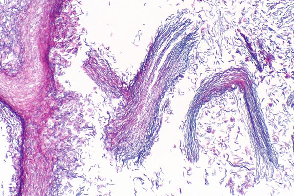
A cholesteatoma is essentially an abnormal accumulation of skin cells in the middle ear, behind the eardrum. While it is not cancerous, it can cause damage to the delicate structures of the ear, leading to hearing loss and other complications. Understanding what a cholesteatoma is and how it develops is crucial for early detection and treatment.
What Happens in the Ear?
The ear is divided into three main sections: the outer ear, the middle ear, and the inner ear. The middle ear contains tiny bones called ossicles, which are responsible for transmitting sound vibrations from the eardrum to the inner ear. A cholesteatoma forms when skin cells grow abnormally in this area, creating a cyst-like structure that can expand over time.
Causes of Cholesteatoma
Several factors can contribute to the development of a cholesteatoma. Understanding these causes can help individuals take preventive measures and seek timely medical care.
Ear Infections
Recurrent or chronic ear infections are one of the most common causes of cholesteatoma. When the eardrum becomes damaged due to repeated infections, it may form a pocket where dead skin cells accumulate. Over time, this pocket can grow and develop into a cholesteatoma.
Eustachian Tube Dysfunction
The Eustachian tube connects the middle ear to the back of the nose and helps regulate air pressure in the ear. If this tube does not function properly, it can lead to negative pressure in the middle ear. This pressure can cause the eardrum to retract, forming a pouch where skin cells can collect and form a cholesteatoma.
Congenital Factors
In some cases, individuals may be born with a cholesteatoma. This type of cholesteatoma is referred to as congenital and occurs when skin cells become trapped in the middle ear during fetal development. Congenital cholesteatomas are typically discovered in childhood during routine ear examinations.
Symptoms of Cholesteatoma
Recognizing the symptoms of a cholesteatoma is essential for early diagnosis and treatment. The signs of this condition can vary depending on its size and location, but there are several common symptoms to watch for.
Hearing Loss
One of the most noticeable symptoms of a cholesteatoma is hearing loss. As the abnormal growth expands, it can damage the ossicles or interfere with their ability to transmit sound. This often results in conductive hearing loss, which affects the ear’s ability to conduct sound waves effectively.
Ear Drainage
- Foul-smelling discharge from the ear
- Persistent drainage that does not resolve with standard treatments
A cholesteatoma can cause persistent ear drainage that often has a foul odor. This discharge occurs because the trapped skin cells within the cyst break down and produce debris, which can leak out of the ear.
Ear Pain and Discomfort
Individuals with a cholesteatoma may experience pain or discomfort in the affected ear. This pain can range from mild to severe and may worsen over time as the cyst grows and presses against surrounding tissues.
Dizziness and Balance Issues
If a cholesteatoma affects the inner ear or damages the structures responsible for balance, it can lead to dizziness or vertigo. These symptoms may make it difficult to maintain balance or perform daily activities.
Diagnosing Cholesteatoma
Accurate diagnosis is critical for effective treatment of a cholesteatoma. Healthcare providers use a combination of physical examinations, imaging tests, and other diagnostic tools to confirm the presence of this condition.
Physical Examination
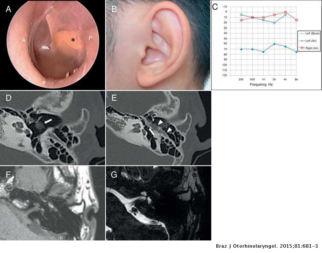
A healthcare provider will begin by examining the ear using an otoscope, a tool that allows them to view the eardrum and middle ear. During this examination, they may notice a retracted eardrum, visible growth, or signs of infection.
Hearing Tests
Hearing tests, such as audiometry, are often performed to assess the extent of hearing loss caused by the cholesteatoma. These tests measure how well the ear can detect different sounds and frequencies.
Imaging Studies
- Computed tomography (CT) scans
- Magnetic resonance imaging (MRI)
Imaging studies like CT scans and MRI are invaluable for diagnosing a cholesteatoma. These tests provide detailed images of the ear’s internal structures, allowing healthcare providers to determine the size and location of the growth.
Treatment Options for Cholesteatoma
Treatment for a cholesteatoma typically involves surgical intervention to remove the abnormal growth and prevent further complications. However, the specific approach depends on the size, location, and severity of the condition.
Surgical Removal
Surgery is the primary treatment for a cholesteatoma. The goal of surgery is to completely remove the growth while preserving as much of the ear’s normal function as possible. There are two main types of surgical procedures used to treat this condition:
- Tympanoplasty: This procedure involves repairing the eardrum and removing the cholesteatoma. It is often combined with ossiculoplasty, which reconstructs or replaces damaged ossicles.
- Mastoidectomy: In more advanced cases, a mastoidectomy may be necessary. This surgery removes the cholesteatoma from the mastoid bone, which is located behind the ear.
Post-Surgical Care
After surgery, patients must follow strict post-operative care instructions to ensure proper healing and reduce the risk of recurrence. This may include regular follow-up appointments, cleaning of the ear, and avoiding activities that could damage the ear.
Non-Surgical Management
In some cases, non-surgical approaches may be used to manage symptoms or prepare for surgery. These may include:
- Antibiotics to treat or prevent infection
- Eardrops to reduce inflammation or drainage
However, it is important to note that non-surgical management is not a cure and should only be used as a temporary measure.
Potential Complications of Untreated Cholesteatoma
If left untreated, a cholesteatoma can lead to serious complications that affect not only the ear but also other parts of the body. Understanding these risks underscores the importance of seeking timely medical care.
Hearing Loss
As the cholesteatoma grows, it can destroy the ossicles or damage the cochlea, leading to permanent hearing loss. This type of hearing loss is often irreversible, even after surgical removal of the growth.
Infections
The trapped debris within a cholesteatoma provides an ideal environment for bacteria to thrive, increasing the risk of recurrent or severe infections. These infections can spread to nearby areas, such as the brain, causing life-threatening complications.
Facial Paralysis
In rare cases, a cholesteatoma can press against the facial nerve, leading to facial paralysis. This occurs when the nerve responsible for controlling facial muscles becomes compressed or damaged.
Meningitis
If the infection spreads to the membranes surrounding the brain and spinal cord, it can cause meningitis. This condition is characterized by fever, headache, and stiffness in the neck and requires immediate medical attention.
Preventing Cholesteatoma
While not all cases of cholesteatoma can be prevented, certain measures can reduce the risk of developing this condition.
Managing Ear Infections
Seeking prompt treatment for ear infections can help prevent damage to the eardrum and reduce the likelihood of cholesteatoma formation. This includes taking prescribed antibiotics and following up with a healthcare provider as needed.
Avoiding Inserting Objects into the Ear
Inserting objects such as cotton swabs or hairpins into the ear can damage the eardrum and increase the risk of cholesteatoma. It is best to clean the outer ear gently and avoid inserting anything into the ear canal.
Monitoring Ear Health
Regular check-ups with an ear specialist can help detect early signs of cholesteatoma, especially in individuals with a history of ear problems or Eustachian tube dysfunction. Early detection improves the chances of successful treatment.


