
A craniopharyngioma, often abbreviated as CP, is a rare and benign brain tumor that develops near the pituitary gland. Despite being non-cancerous, this type of tumor can significantly impact a person’s health due to its location and potential to interfere with critical brain functions. In this article, we will explore the intricacies of this condition, including its causes, symptoms, diagnosis, and treatment options.
What Is Craniopharyngioma?
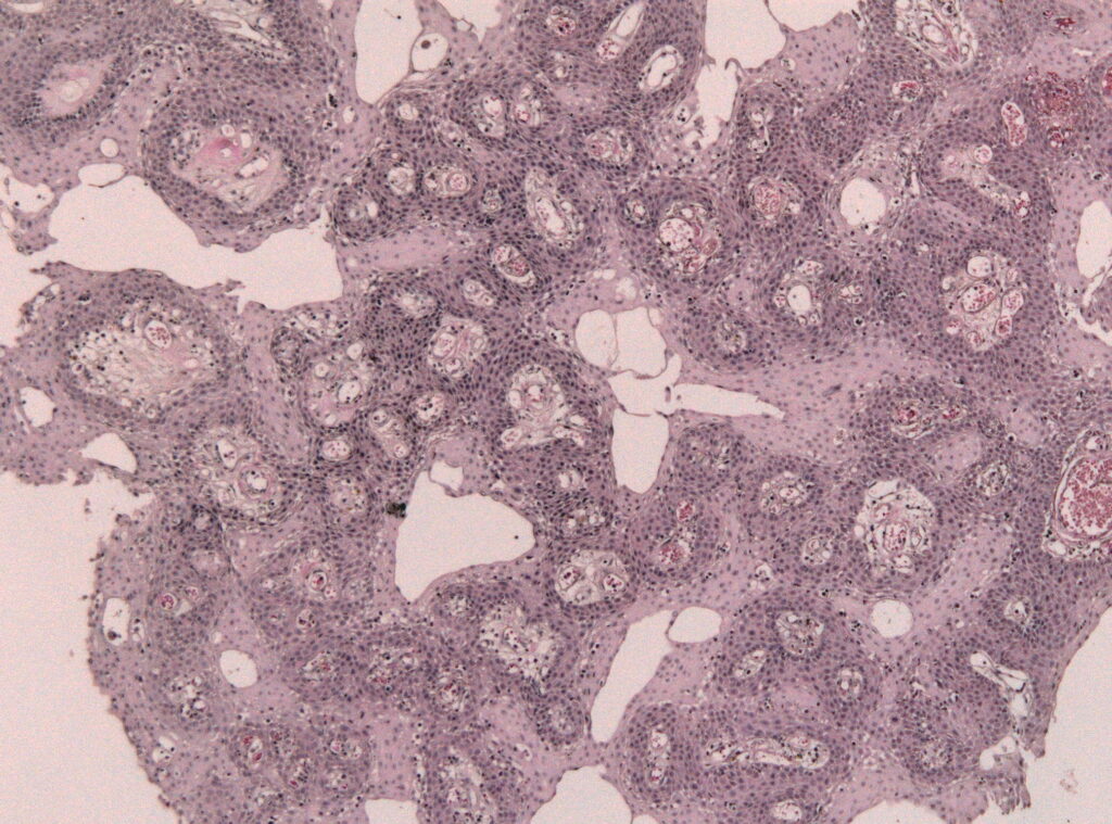
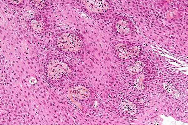
Craniopharyngioma is a slow-growing tumor that typically arises in the area of the brain near the pituitary gland, which is responsible for regulating hormones in the body. These tumors originate from remnants of cells that played a role in the development of the pituitary gland during fetal growth. Although they are classified as benign, their proximity to vital structures such as the hypothalamus and optic nerves makes them potentially life-altering.
Key Characteristics of Craniopharyngioma
- They are most commonly diagnosed in children and adults over the age of fifty.
- These tumors tend to grow slowly but can cause significant damage by pressing on surrounding tissues.
- Craniopharyngiomas can be either solid or cystic, with some cases presenting a combination of both.
Causes and Risk Factors
The exact cause of craniopharyngioma remains unknown. However, researchers believe that these tumors arise from embryonic cells that were involved in the formation of the pituitary gland during early fetal development. These cells may remain dormant for years before eventually forming a tumor.
There are no known risk factors associated with craniopharyngioma. Unlike many other types of tumors, it does not appear to be linked to genetic predispositions or environmental factors. This unpredictability makes it challenging to identify individuals who may be at higher risk.
Symptoms of Craniopharyngioma
The symptoms of craniopharyngioma vary depending on the size and location of the tumor. Because the tumor grows near the pituitary gland and hypothalamus, it can disrupt hormone production and affect nearby structures such as the optic nerves. Common symptoms include:
Hormonal Imbalances
- Growth delays in children
- Delayed puberty
- Fatigue and weakness
- Increased thirst and urination (diabetes insipidus)
- Weight gain or loss
Neurological Symptoms
- Vision problems, such as blurred vision or partial blindness
- Headaches
- Nausea and vomiting
- Difficulty maintaining balance
Cognitive and Emotional Changes
- Memory problems
- Mood swings
- Behavioral changes
Diagnosis of Craniopharyngioma
Diagnosing craniopharyngioma requires a combination of clinical evaluation, imaging studies, and laboratory tests. Early detection is crucial to prevent complications and improve treatment outcomes.
Medical History and Physical Examination
A healthcare provider will begin by reviewing the patient’s medical history and conducting a thorough physical examination. During this process, they may inquire about symptoms such as headaches, vision changes, or hormonal irregularities.
Imaging Studies
Imaging plays a pivotal role in diagnosing craniopharyngioma. The two primary imaging techniques used are:
- Magnetic Resonance Imaging (MRI): MRI provides detailed images of the brain and is particularly effective in identifying tumors near the pituitary gland. It helps determine the size, shape, and exact location of the tumor.
- Computed Tomography (CT) Scan: CT scans use X-rays to create cross-sectional images of the brain. They are especially useful for detecting calcifications, which are often present in craniopharyngiomas.
Hormone Testing
Blood tests may be performed to assess hormone levels. Since the pituitary gland controls various hormones, abnormalities in hormone production can indicate the presence of a tumor. Tests may include measurements of thyroid-stimulating hormone, cortisol, growth hormone, and others.
Visual Field Testing
Given that craniopharyngiomas can compress the optic nerves, visual field testing is often conducted to evaluate any loss of peripheral vision or other visual impairments.
Treatment Options for Craniopharyngioma
Treating craniopharyngioma involves a multidisciplinary approach, combining surgery, radiation therapy, and medication management. The choice of treatment depends on factors such as the size and location of the tumor, the patient’s age, and overall health.
Surgical Removal
Surgery is typically the first line of treatment for craniopharyngioma. The goal is to remove as much of the tumor as possible while minimizing damage to surrounding tissues. There are two main surgical approaches:
- Transsphenoidal Surgery: This minimally invasive procedure involves accessing the tumor through the nose and sphenoid sinus. It is often preferred for smaller tumors located near the pituitary gland.
- Craniotomy: For larger tumors or those extending into other areas of the brain, a craniotomy may be necessary. This involves opening the skull to access and remove the tumor.
While surgery can effectively reduce the size of the tumor, complete removal is not always possible due to the tumor’s proximity to critical structures.
Radiation Therapy
In cases where the tumor cannot be fully removed or if it recurs after surgery, radiation therapy may be recommended. Radiation uses high-energy beams to target and destroy tumor cells. Types of radiation therapy include:
- External Beam Radiation: Delivers precise doses of radiation to the tumor site.
- Stereotactic Radiosurgery: A highly focused form of radiation that targets the tumor with minimal impact on surrounding tissues.
Hormone Replacement Therapy
Since craniopharyngiomas often disrupt normal hormone production, patients may require lifelong hormone replacement therapy. This treatment involves administering synthetic versions of hormones that the body is no longer producing adequately. Examples include:
- Thyroid hormone
- Cortisol
- Growth hormone
- Sex hormones
Monitoring and Follow-Up Care
Regular follow-up appointments are essential to monitor the patient’s recovery and detect any recurrence of the tumor. Follow-up care may involve periodic imaging studies, hormone level assessments, and evaluations of neurological function.
Living with Craniopharyngioma
Managing craniopharyngioma extends beyond medical treatments. Patients and their families must navigate the emotional, social, and practical challenges that come with living with this condition.
Emotional Support
Dealing with a brain tumor can be overwhelming. Counseling, support groups, and mental health services can provide valuable emotional support for patients and their loved ones.
Lifestyle Adjustments
Depending on the severity of symptoms and treatment side effects, patients may need to make certain lifestyle adjustments. These could include dietary changes, modifications to daily routines, or adaptations to accommodate physical limitations.
Educational and Occupational Considerations
Children diagnosed with craniopharyngioma may face challenges in school due to cognitive or developmental delays. Adults may need workplace accommodations or vocational rehabilitation services to continue working effectively.
Advances in Research and Treatment
Ongoing research is paving the way for improved understanding and treatment of craniopharyngioma. Scientists are exploring new surgical techniques, advanced radiation therapies, and targeted drug treatments to enhance outcomes for patients.
Targeted Therapies
Recent advancements in molecular biology have led to the identification of specific genetic mutations associated with craniopharyngioma. This knowledge opens the door to developing targeted therapies that can more effectively treat the tumor while sparing healthy tissue.
Minimally Invasive Procedures
Technological innovations in neurosurgery are enabling surgeons to perform increasingly precise and less invasive procedures. These advancements aim to reduce recovery times and minimize complications for patients undergoing treatment.


