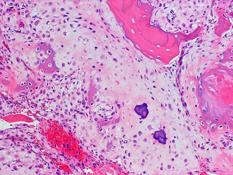
Chondrosarcoma, often abbreviated as CS, is a rare and complex form of bone cancer that originates in the cartilage cells. This type of cancer primarily affects adults and is known for its slow-growing nature, although certain subtypes can be aggressive. Unlike other forms of bone cancer, chondrosarcoma poses unique challenges in diagnosis and treatment due to its resistance to conventional therapies such as chemotherapy and radiation. In this article, we will explore the intricacies of this condition, including its causes, symptoms, diagnostic methods, treatment options, and ongoing research.

Understanding Chondrosarcoma
Chondrosarcoma is a malignant tumor that arises from cartilage, the flexible connective tissue found in various parts of the body, including joints, ribs, and the spine. Cartilage plays a crucial role in providing structural support and cushioning between bones. When cancer develops in this tissue, it disrupts normal cellular processes and leads to the formation of abnormal growths.
This type of cancer typically occurs in adults between the ages of 40 and 70, with a higher prevalence in men compared to women. It accounts for approximately 20 percent of all primary bone cancers, making it the second most common type after osteosarcoma. While chondrosarcoma can develop in any bone containing cartilage, it is most commonly found in the pelvis, femur, humerus, and ribs.
Types of Chondrosarcoma
There are several subtypes of chondrosarcoma, each with distinct characteristics and levels of aggressiveness:
- Conventional Chondrosarcoma: This is the most common subtype, accounting for about 85 percent of cases. It is generally slow-growing and less likely to metastasize.
- Dedifferentiated Chondrosarcoma: This aggressive subtype combines conventional chondrosarcoma with high-grade sarcoma, making it more challenging to treat.
- Mesenchymal Chondrosarcoma: A rare and fast-growing variant that often affects younger individuals and tends to spread to other parts of the body.
- Clear Cell Chondrosarcoma: A slow-growing subtype that typically occurs in the ends of long bones, such as the femur or humerus.
Causes and Risk Factors
The exact cause of chondrosarcoma remains unknown, but researchers have identified several factors that may increase the risk of developing this condition:
Genetic Predisposition
Some inherited genetic disorders are associated with an increased risk of chondrosarcoma. These include:
- Ollier Disease: A rare condition characterized by the development of multiple benign cartilage tumors, known as enchondromas, which can transform into malignant chondrosarcomas.
- Maffucci Syndrome: Similar to Ollier disease, this condition involves the presence of enchondromas along with benign blood vessel tumors called hemangiomas.
Radiation Exposure
Prior exposure to radiation therapy, particularly for the treatment of other cancers, has been linked to an elevated risk of developing chondrosarcoma. The latency period between radiation exposure and the onset of cancer can range from several years to decades.
Age and Gender
As mentioned earlier, chondrosarcoma predominantly affects adults over the age of 40, with a higher incidence in males. The reasons for this gender disparity are not fully understood but may be related to hormonal or genetic factors.
Symptoms of Chondrosarcoma
The symptoms of chondrosarcoma can vary depending on the location and size of the tumor. However, some common signs and symptoms include:
- Persistent pain in the affected area, which may worsen at night or with physical activity.
- Swelling or a noticeable lump near the tumor site.
- Limited range of motion if the tumor is located near a joint.
- Fatigue or general weakness, especially in advanced stages.
It is important to note that these symptoms are nonspecific and can mimic other conditions, such as arthritis or benign bone tumors. As a result, chondrosarcoma is often misdiagnosed or detected at a later stage.
Diagnosis of Chondrosarcoma
Diagnosing chondrosarcoma requires a comprehensive approach that includes clinical evaluation, imaging studies, and biopsy. Early and accurate diagnosis is critical for determining the appropriate treatment plan.
Imaging Studies
Imaging techniques play a pivotal role in identifying and characterizing chondrosarcoma. Commonly used methods include:
- X-rays: These provide initial insights into bone abnormalities, such as calcifications or lesions, which may suggest the presence of a tumor.
- Magnetic Resonance Imaging (MRI): MRI offers detailed images of soft tissues and helps assess the extent of tumor involvement.
- Computed Tomography (CT) Scans: CT scans are useful for evaluating bone destruction and planning surgical interventions.
- Positron Emission Tomography (PET) Scans: PET scans can help determine whether the cancer has spread to other parts of the body.
Biopsy
A biopsy is essential for confirming the diagnosis of chondrosarcoma. During this procedure, a small sample of tissue is extracted from the tumor and examined under a microscope. There are two main types of biopsies:
- Needle Biopsy: A thin needle is used to remove tissue from the tumor. This method is minimally invasive and often preferred for initial diagnosis.
- Surgical Biopsy: In some cases, a larger sample may be required, necessitating a surgical procedure.
Treatment Options for Chondrosarcoma
The treatment of chondrosarcoma depends on several factors, including the tumor’s size, location, grade, and whether it has metastasized. Due to its resistance to chemotherapy and radiation, surgery remains the cornerstone of treatment.
Surgical Intervention
Surgery is the primary treatment for chondrosarcoma and aims to remove the tumor while preserving as much function as possible. The specific surgical approach depends on the tumor’s characteristics:
- Wide Local Excision: This involves removing the tumor along with a margin of healthy tissue to reduce the risk of recurrence.
- Limb-Sparing Surgery: In cases where the tumor is located in an extremity, surgeons may opt to remove the tumor while preserving the limb’s functionality.
- Amputation: Although less common today, amputation may be necessary if the tumor is large or located in a critical area where limb preservation is not feasible.
Radiation Therapy
Radiation therapy is generally not effective for treating conventional chondrosarcoma due to the tumor’s low sensitivity to radiation. However, it may be used in cases where surgery is not possible or for managing symptoms in advanced stages.
Chemotherapy
Similar to radiation therapy, chemotherapy has limited efficacy in treating chondrosarcoma. It is occasionally used for aggressive subtypes, such as mesenchymal or dedifferentiated chondrosarcoma, which may respond better to systemic treatments.
Targeted Therapy and Emerging Treatments
Recent advances in cancer research have led to the development of targeted therapies that focus on specific molecular pathways involved in tumor growth. While still in the experimental stages, these treatments hold promise for improving outcomes in patients with chondrosarcoma.
Ongoing Research and Future Directions
Despite significant progress in understanding chondrosarcoma, many challenges remain, particularly in terms of treatment options for advanced or metastatic cases. Researchers are actively exploring new avenues to enhance diagnosis and therapy:
Genomic Studies
Genomic profiling of chondrosarcoma tumors has revealed mutations in genes such as IDH1 and IDH2, which play a role in cellular metabolism. Targeting these mutations could lead to the development of novel therapies tailored to individual patients.
Immunotherapy
Immunotherapy, which harnesses the body’s immune system to fight cancer, is another area of interest. Early studies suggest that immune checkpoint inhibitors may have potential applications in treating chondrosarcoma.
Collaborative Efforts
International collaborations and clinical trials are essential for advancing knowledge and improving patient outcomes. By pooling resources and expertise, researchers aim to accelerate the discovery of effective treatments for this challenging disease.


