
Teratomas, often abbreviated as TM, are a unique type of tumor that originates from pluripotent cells, which have the ability to develop into various types of tissues. These tumors can contain components derived from all three germ layers: ectoderm, mesoderm, and endoderm. This characteristic makes them distinct from other types of tumors and gives rise to their complex nature. In this article, we will delve into the details of teratomas, focusing on two primary categories: mature and immature teratomas.
Understanding Teratomas
Teratomas are fascinating because they can include a wide variety of tissue types, such as hair, teeth, bone, and even more complex structures like organs or glandular tissue. They are most commonly found in the ovaries and testes but can also occur in other areas of the body, including the sacrococcygeal region, mediastinum, and central nervous system.
Origins of Teratomas
The development of teratomas is linked to the presence of pluripotent cells, which are capable of differentiating into multiple cell types. These cells are typically present during early embryonic development, and their abnormal proliferation can lead to the formation of teratomas. The exact cause of this abnormal growth is not fully understood, but genetic and environmental factors may play a role.
Germ Layers and Their Contribution
- Ectoderm: This layer contributes to the formation of skin, hair, nails, and nervous tissue. In teratomas, ectodermal components may manifest as skin-like structures or neural tissue.
- Mesoderm: Responsible for forming muscle, bone, cartilage, and connective tissue, the mesoderm can give rise to bony structures or muscular elements within teratomas.
- Endoderm: This layer forms the lining of internal organs and glands. Endodermal contributions to teratomas might include glandular tissue or respiratory epithelium.
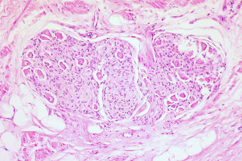
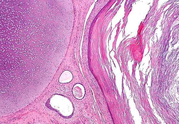
Mature Teratomas
Mature teratomas are the most common type of teratoma and are generally benign. They are composed of well-differentiated tissues that resemble normal tissues found in the body. Despite their benign nature, these tumors can still cause complications depending on their size and location.
Characteristics of Mature Teratomas
Mature teratomas are often referred to as dermoid cysts when they occur in the ovaries. They are encapsulated and typically contain a mixture of tissues, such as skin, hair follicles, sebaceous glands, and sometimes even teeth or bone. These tumors are slow-growing and rarely spread to other parts of the body.
Common Locations
- Ovaries: The majority of mature teratomas are found in the ovaries, particularly in women of reproductive age.
- Testes: In males, these tumors can occur in the testes, although they are less common than in females.
- Sacrococcygeal Region: This is the most frequent site of teratomas in infants and children.
Diagnosis and Treatment
Diagnosing mature teratomas usually involves imaging studies such as ultrasound, computed tomography, or magnetic resonance imaging. These imaging techniques help identify the presence of solid and cystic components within the tumor. Surgical removal is the standard treatment for mature teratomas, and the prognosis is generally excellent after complete excision.
Immature Teratomas
Immature teratomas are less common than mature teratomas and are considered malignant. These tumors contain poorly differentiated or embryonal tissues, which can include primitive neural tissue. The presence of these immature elements increases the risk of aggressive behavior and metastasis.
Characteristics of Immature Teratomas
Immature teratomas are typically found in younger patients, with a higher incidence in adolescents and young adults. Unlike mature teratomas, these tumors are not encapsulated and may invade surrounding tissues. The degree of immaturity is graded based on the proportion of immature tissue present, with higher grades indicating a greater likelihood of malignancy.
Grading System
- Grade 1: Contains mostly mature tissue with minimal immature elements.
- Grade 2: Has a moderate amount of immature tissue.
- Grade 3: Predominantly composed of immature tissue, indicating a high risk of malignancy.
Common Locations
Immature teratomas are most frequently found in the ovaries, but they can also occur in the testes, mediastinum, and central nervous system. The location of the tumor can influence its clinical presentation and treatment approach.
Diagnosis and Treatment
Diagnosing immature teratomas requires a combination of imaging studies and biopsy. The biopsy helps determine the grade of the tumor by examining the proportion of immature tissue. Treatment typically involves surgical removal followed by chemotherapy, especially for higher-grade tumors. The prognosis depends on the grade of the tumor and whether it has spread to other parts of the body.
Clinical Presentation and Symptoms
The symptoms of teratomas vary depending on their location and size. In some cases, these tumors may be asymptomatic and discovered incidentally during routine examinations or imaging studies. However, larger tumors or those located in specific areas can cause noticeable symptoms.
Symptoms Based on Location
- Ovarian Teratomas: May cause abdominal pain, bloating, or irregular menstrual cycles.
- Testicular Teratomas: Often present as a painless lump or swelling in the testicle.
- Sacrococcygeal Teratomas: Can cause visible swelling at the base of the spine and may interfere with bowel or bladder function.
- Mediastinal Teratomas: May lead to chest pain, coughing, or difficulty breathing if they press on nearby structures.
Pathophysiology of Teratomas
The pathophysiology of teratomas is rooted in the abnormal proliferation of pluripotent cells. These cells retain the ability to differentiate into various tissue types, leading to the formation of tumors that contain a diverse array of structures. The exact mechanisms that trigger this abnormal growth are not fully understood, but several theories have been proposed.
Theories of Development
- Parthenogenetic Activation: Suggests that teratomas arise from unfertilized eggs that undergo spontaneous division and differentiation.
- Embryonic Cell Migration: Proposes that teratomas result from the abnormal migration of embryonic cells during early development.
- Genetic Mutations: Certain genetic abnormalities may predispose individuals to the development of teratomas.
Risk Factors and Prevention
While the exact causes of teratomas remain unclear, certain risk factors have been identified. These include genetic predispositions, environmental influences, and hormonal imbalances. Unfortunately, there are no definitive preventive measures for teratomas, as their development is often unpredictable.
Potential Risk Factors
- Genetic Syndromes: Conditions such as Turner syndrome or Klinefelter syndrome may increase the risk of developing teratomas.
- Hormonal Factors: Fluctuations in hormone levels, particularly during puberty or pregnancy, may contribute to the development of ovarian teratomas.
- Environmental Exposures: Exposure to certain chemicals or radiation may play a role in the formation of these tumors.
Complications Associated with Teratomas
Both mature and immature teratomas can lead to complications, although the nature and severity of these complications differ. Understanding these potential issues is crucial for managing the condition effectively.
Complications of Mature Teratomas
- Torsion: Large ovarian teratomas can cause the ovary to twist, leading to severe pain and reduced blood flow.
- Rupture: The tumor may rupture, causing spillage of its contents into the abdominal cavity and leading to inflammation or infection.
- Compression: Depending on their size and location, teratomas can compress nearby organs or structures, causing functional impairment.
Complications of Immature Teratomas
- Metastasis: Higher-grade immature teratomas have the potential to spread to other parts of the body, complicating treatment.
- Recurrence: Even after surgical removal, immature teratomas may recur, necessitating long-term monitoring.
- Chemotherapy Side Effects: The use of chemotherapy to treat malignant teratomas can lead to significant side effects, including nausea, fatigue, and immune suppression.
Future Directions in Research
Research into teratomas continues to evolve, with scientists exploring new methods for diagnosis, treatment, and prevention. Advances in molecular biology and genetics hold promise for better understanding the underlying causes of these tumors and developing targeted therapies.
Potential Areas of Focus
- Genomic Studies: Investigating the genetic mutations associated with teratomas may reveal new biomarkers for early detection.
- Targeted Therapies: Developing treatments that specifically target the pluripotent cells responsible for teratoma formation could improve outcomes.
- Immunotherapy: Harnessing the immune system to combat malignant teratomas is an emerging area of interest.


