
Liposarcoma, often abbreviated as LPS, is a rare form of cancer that arises in the fat cells of the body. It is classified as a type of soft tissue sarcoma, which means it develops in the connective tissues that support and surround other structures in the body. Liposarcoma can occur anywhere in the body but is most commonly found in the abdomen, limbs, or areas behind the abdominal cavity known as the retroperitoneum. This article delves into the various types of liposarcoma, its potential causes, methods of diagnosis, and the treatment options available to patients.
Understanding Liposarcoma
Liposarcoma is a complex disease that affects people of all ages, though it is more frequently diagnosed in adults between the ages of fifty and seventy. Despite being a rare condition, understanding its nature is crucial for early detection and effective management. The tumors associated with this condition are typically slow-growing, which can make them difficult to detect in their early stages. However, they can grow to significant sizes if left untreated, leading to complications depending on their location.
Types of Liposarcoma
Liposarcoma is categorized into several distinct types based on its cellular characteristics and behavior. Each type has unique features that influence its prognosis and treatment approach. Below are the primary classifications:
Well-Differentiated Liposarcoma
- This is the most common type of liposarcoma and tends to grow slowly.
- It resembles normal fat cells under a microscope but contains additional genetic material that distinguishes it from benign fatty tumors.
- Although it is less likely to spread to other parts of the body, it can recur locally if not completely removed.
Dedifferentiated Liposarcoma
- This type begins as a well-differentiated liposarcoma but transforms into a more aggressive form over time.
- The dedifferentiated areas lack the typical fat cell appearance and may behave more like high-grade sarcomas.
- It poses a higher risk of spreading to other organs compared to well-differentiated liposarcoma.
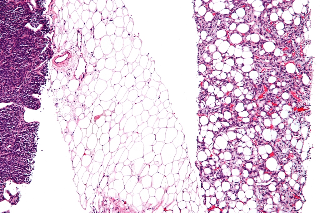
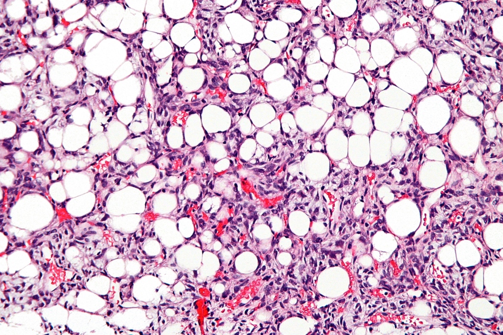
Myxoid Liposarcoma
- Characterized by a gelatinous appearance due to the presence of mucous-like material within the tumor.
- It often occurs in the limbs and has a moderate likelihood of metastasis, particularly to unusual sites such as the bones or lungs.
- Treatment outcomes are generally favorable when detected early.
Pleomorphic Liposarcoma
- This is one of the rarest and most aggressive forms of liposarcoma.
- It consists of highly irregular and large tumor cells, making it challenging to treat.
- Pleomorphic liposarcoma has a higher tendency to metastasize and requires aggressive treatment strategies.
Mixed-Type Liposarcoma
- As the name suggests, this type exhibits characteristics of multiple subtypes of liposarcoma.
- Its behavior and prognosis depend on the predominant subtype present within the tumor.
Potential Causes of Liposarcoma
The exact cause of liposarcoma remains unknown, but researchers have identified certain factors that may contribute to its development. These include:
Genetic Mutations
- Changes in specific genes, such as those involved in regulating cell growth and division, are believed to play a role in the formation of liposarcoma.
- For instance, abnormalities in chromosomes thirteen and eighteen are commonly observed in certain types of liposarcoma.
Radiation Exposure
- Prior exposure to radiation therapy for other cancers has been linked to an increased risk of developing liposarcoma later in life.
- The latency period between radiation exposure and tumor development can span several years.
Chemical Exposures
- Some studies suggest that exposure to certain chemicals, such as vinyl chloride or dioxins, might increase the risk of soft tissue sarcomas, including liposarcoma.
- However, definitive evidence linking these substances to liposarcoma is still limited.
Other Risk Factors
- Age is a significant factor, as liposarcoma is more prevalent in older adults.
- A family history of cancer or inherited syndromes associated with soft tissue tumors may also elevate the risk.
Diagnosing Liposarcoma
Diagnosing liposarcoma involves a combination of clinical evaluation, imaging studies, and laboratory tests. Early and accurate diagnosis is essential for determining the appropriate treatment plan. Here are the key steps involved in diagnosing this condition:
Clinical Evaluation
- A healthcare provider will begin by taking a detailed medical history and performing a physical examination.
- They will assess the size, location, and mobility of any suspicious lumps or masses.
Imaging Studies
- Magnetic Resonance Imaging (MRI): MRI scans provide detailed images of soft tissues and are particularly useful for identifying the extent of the tumor.
- Computed Tomography (CT) Scan: CT scans help visualize internal structures and determine whether the tumor has spread to nearby organs or lymph nodes.
- Ultrasound: This non-invasive technique uses sound waves to create images of the affected area and is often used during initial evaluations.
Bioopsy
- A biopsy involves removing a small sample of tissue from the tumor for microscopic examination.
- This procedure helps confirm the diagnosis and classify the specific type of liposarcoma.
- Biopsies can be performed using needles or through surgical excision, depending on the tumor’s location and accessibility.
Laboratory Tests
- Blood tests may be conducted to rule out other conditions and assess overall health before initiating treatment.
- While there are no specific blood markers for liposarcoma, these tests provide valuable information about liver and kidney function, which is important for planning therapies.
Treatment Options for Liposarcoma
The treatment of liposarcoma depends on several factors, including the type of tumor, its size and location, and the patient’s overall health. A multidisciplinary team of specialists, including oncologists, surgeons, and radiologists, typically collaborates to develop a personalized treatment plan. Below are the main treatment modalities:
Surgery
- Surgical removal of the tumor is the primary treatment for most cases of liposarcoma.
- The goal is to remove the entire tumor along with a margin of healthy tissue to reduce the risk of recurrence.
- In cases where the tumor is located in critical areas, such as near vital organs, achieving complete resection can be challenging.
Radiation Therapy
- Radiation therapy uses high-energy beams to target and destroy cancer cells.
- It is often used before surgery to shrink large tumors or after surgery to eliminate any remaining cancer cells.
- Advanced techniques, such as intensity-modulated radiation therapy, minimize damage to surrounding healthy tissues.
Chemotherapy
- Chemotherapy involves the use of drugs to kill cancer cells or stop them from growing.
- It is typically reserved for advanced or metastatic cases of liposarcoma that cannot be treated with surgery alone.
- Newer chemotherapy agents and combinations are continually being researched to improve outcomes.
Targeted Therapy
- Targeted therapies focus on specific molecular pathways involved in the growth and survival of cancer cells.
- Drugs such as pazopanib have shown promise in treating certain types of liposarcoma by inhibiting angiogenesis, the process by which tumors form new blood vessels.
- These treatments are often considered when traditional therapies are ineffective.
Clinical Trials
- Participating in clinical trials provides access to cutting-edge treatments that are not yet widely available.
- Trials may involve novel drugs, immunotherapies, or innovative surgical techniques aimed at improving survival rates and quality of life.
- Patients should discuss eligibility and potential benefits with their healthcare providers.
Supportive Care
- Beyond active treatment, supportive care plays a vital role in managing symptoms and side effects.
- Physical therapy, pain management, and psychological counseling can help patients cope with the physical and emotional challenges of living with liposarcoma.
- Nutritional support and rehabilitation services further enhance recovery and overall well-being.


