
Legg-Calve-Perthes Disease, often abbreviated as LCPD, is a rare childhood condition that affects the hip joint. This disorder occurs when the blood supply to the ball-shaped head of the thigh bone, also known as the femoral head, is temporarily disrupted. Without adequate blood flow, the bone tissue begins to deteriorate, leading to potential deformities and long-term complications if not managed properly. In this article, we will explore the intricacies of this condition, including its symptoms, diagnostic methods, and available care options.
Understanding Legg-Calve-Perthes Disease
Legg-Calve-Perthes Disease primarily affects children between the ages of four and eight, though it can occur in younger or older children as well. It is more commonly observed in boys than in girls. The exact cause of the disease remains unknown, but researchers believe that genetic factors, trauma, and abnormal blood clotting may play a role in its development.
The hip joint is a ball-and-socket joint where the rounded head of the femur fits into the cup-shaped socket of the pelvis. When the blood supply to the femoral head is interrupted, the bone cells begin to die, resulting in a condition called avascular necrosis. Over time, the body attempts to repair the damaged bone by replacing it with new bone tissue. However, this regenerative process can lead to an irregularly shaped femoral head, which may cause pain and restricted movement in the affected hip.
Risk Factors for Developing the Condition
- Age: Children between the ages of four and eight are most susceptible to developing the disease.
- Gender: Boys are more likely to be diagnosed with the condition than girls.
- Family History: A family history of the disease increases the likelihood of a child developing it.
- Delayed Bone Growth: Children who experience delayed skeletal maturity may have a higher risk.
Symptoms of Legg-Calve-Perthes Disease
The symptoms of this condition can vary depending on the stage of the disease and the severity of the blood supply disruption. Some children may experience mild discomfort, while others may face significant pain and mobility issues. Below are the most common symptoms associated with the disease:
Early Warning Signs
- Limping: One of the earliest signs of the disease is a noticeable limp, often described as painless at first. The limp occurs because the affected leg may appear shorter due to the irregular shape of the femoral head.
- Hip Pain: As the disease progresses, children may begin to experience pain in the hip, groin, or knee. The pain may worsen with physical activity and improve with rest.
- Reduced Range of Motion: Parents may notice that their child has difficulty moving the affected leg. Activities such as running, jumping, or even sitting cross-legged may become challenging.
Advanced Symptoms
- Muscle Weakness: The muscles surrounding the hip joint may weaken over time due to reduced use and discomfort.
- Joint Stiffness: As the femoral head begins to reshape, stiffness in the hip joint may become more pronounced.
- Pain During Physical Activity: Children may avoid participating in sports or other activities due to increasing pain and discomfort.
Diagnosing the Condition
Early diagnosis of Legg-Calve-Perthes Disease is crucial for effective management and treatment. Since the symptoms can mimic other conditions, such as juvenile arthritis or a hip injury, a thorough evaluation by a healthcare professional is essential. Below are the primary methods used to diagnose the condition:
Physical Examination
A doctor will begin by conducting a comprehensive physical examination. During this assessment, they will observe the child’s gait and check for any signs of limping. They will also evaluate the range of motion in the hip joint by asking the child to perform specific movements, such as bending or rotating the leg. Limited mobility and discomfort during these exercises may indicate the presence of the disease.
Imaging Tests
To confirm the diagnosis, imaging tests are typically performed. These tests provide detailed images of the hip joint and help determine the extent of damage to the femoral head.
- X-rays: X-rays are the most common imaging tool used to diagnose the condition. They can reveal changes in the shape and density of the femoral head, as well as any signs of bone deterioration.
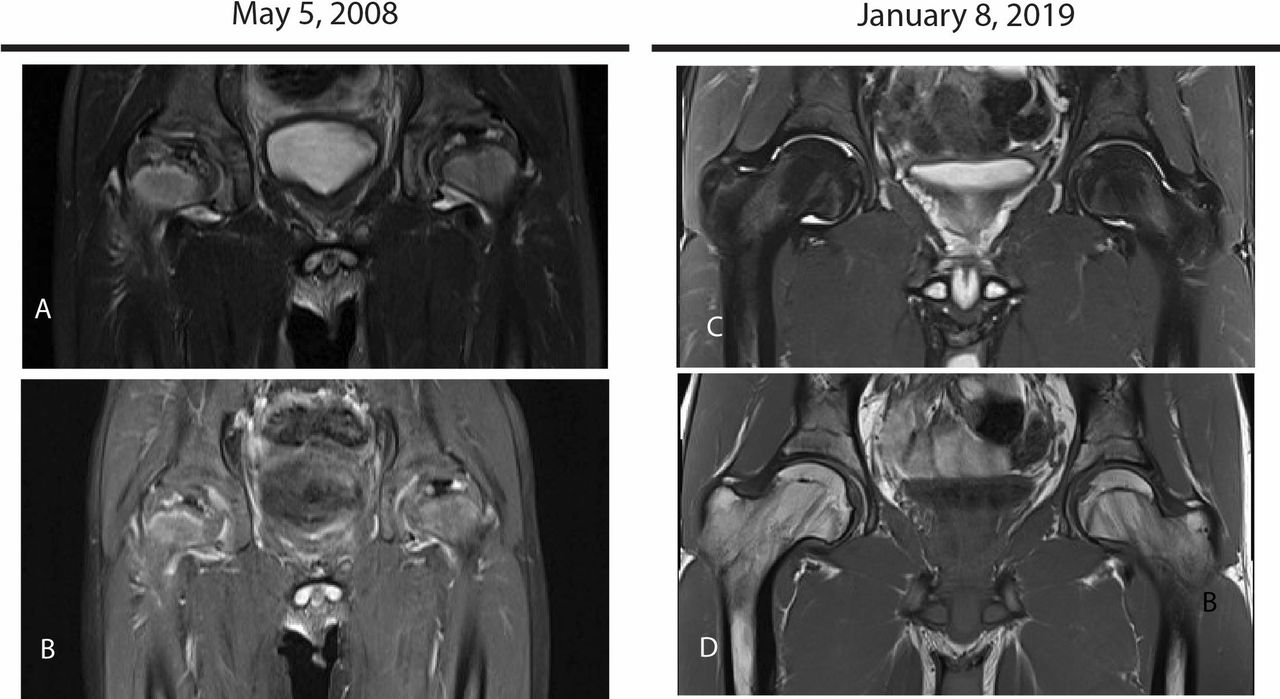
- Magnetic Resonance Imaging (MRI): An MRI provides a more detailed view of the soft tissues and bone marrow. This test is particularly useful for detecting early-stage avascular necrosis before it becomes visible on an X-ray.
- Bone Scans: A bone scan involves injecting a small amount of radioactive material into the bloodstream. The material accumulates in areas of the bone with active healing, helping to identify regions affected by the disease.
Treatment Options for Managing the Condition
The goal of treatment for Legg-Calve-Perthes Disease is to preserve the shape of the femoral head and maintain proper alignment within the hip joint. Treatment plans are tailored to the child’s age, the severity of the condition, and the stage of the disease. Below are the most common approaches to managing the condition:
Non-Surgical Treatments
In mild cases, non-surgical interventions may be sufficient to manage symptoms and promote healing. These treatments focus on reducing pain, improving mobility, and supporting the natural regeneration of the femoral head.
- Rest and Activity Modification: Limiting high-impact activities, such as running and jumping, can help reduce stress on the hip joint. Encouraging low-impact exercises, such as swimming, may also aid in maintaining muscle strength without aggravating the condition.
- Physical Therapy: A physical therapist can design a customized exercise program to improve flexibility, strengthen the surrounding muscles, and enhance joint stability.
- Orthopedic Devices: In some cases, braces or casts may be used to keep the femoral head properly aligned within the hip socket. These devices help ensure that the bone heals in the correct position.
Surgical Interventions
If the condition is severe or does not respond to non-surgical treatments, surgery may be necessary. Surgical procedures aim to restore the shape of the femoral head and improve joint function.
- Osteotomy: This procedure involves cutting and repositioning the bones around the hip joint to improve alignment. By redistributing weight-bearing forces, an osteotomy can help prevent further damage to the femoral head.
- Femoral Head Reshaping: In advanced cases, surgeons may reshape the femoral head to fit better within the hip socket. This technique helps reduce pain and improve mobility.
Long-Term Care and Monitoring
Children diagnosed with this condition require ongoing care and monitoring to ensure proper healing and minimize the risk of complications. Regular follow-up appointments with an orthopedic specialist are essential to track the progress of the disease and make any necessary adjustments to the treatment plan.
Preventing Complications
Without appropriate treatment, Legg-Calve-Perthes Disease can lead to long-term complications, such as arthritis and chronic hip pain. Early intervention and adherence to the prescribed treatment plan are key to preventing these outcomes. Parents should encourage their child to follow all medical recommendations and report any new symptoms promptly.
Emotional and Psychological Support
Living with a chronic condition can be challenging for both the child and their family. Emotional support from parents, teachers, and healthcare providers plays a vital role in helping the child cope with the physical and emotional aspects of the disease. Counseling or support groups may also be beneficial for addressing any anxiety or frustration related to the condition.
Conclusion
Legg-Calve-Perthes Disease is a complex condition that requires careful attention and management. By understanding the symptoms, seeking timely diagnosis, and following a comprehensive treatment plan, children with this condition can achieve optimal outcomes and lead active, fulfilling lives.


